

2026 Nobel Prize in Medicine Honors Optogenetics, Controlling Brain Cells With Light
The prize goes to Karl Deisseroth, Peter Hegemann and Georg Nagel for their contributions to light-sensitive proteins and the development of optogenetic methods.
· 3 min read
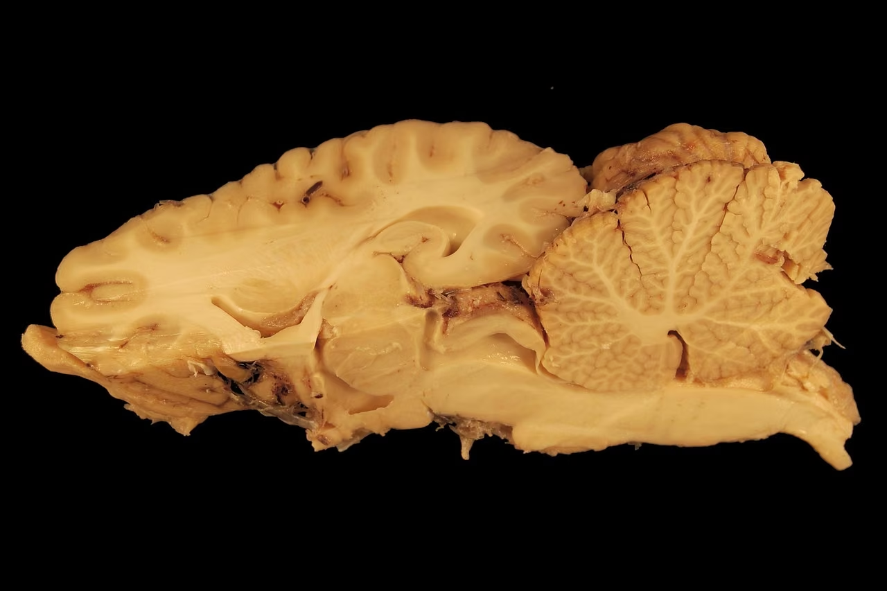
The 2026 Nobel Prize in Medicine was awarded to Karl Deisseroth, Peter Hegemann and Georg Nagel for their work on light-sensitive proteins and their contributions to developing optogenetic techniques. The method lets scientists not only observe specific nerve cells but also control them with light.
From a single-celled alga to neurons
The technology's origin lies not in treating a disease but in the question of how a single-celled organism perceives light. Peter Hegemann of Humboldt University of Berlin and Georg Nagel of the University of Würzburg studied how the single-celled alga Chlamydomonas responds to light. Although it has no eyes, the organism can sense light and adjust its movement to its surroundings.
The researchers discovered channelrhodopsin, a protein in the alga's cell membrane. When light reaches the light-sensitive molecule in the protein, its structure changes and a channel opens that lets positively charged ions pass through the membrane. This ion movement creates the signal that makes the alga respond to light.
The discovery gained importance because neurons also regulate their activity through ion movement across membranes. If light-sensitive proteins could be placed in neurons, those cells could be activated in a controlled way with light. Karl Deisseroth, a psychiatrist and bioengineering researcher at Stanford University in the US, and his colleagues helped develop the field in this direction.
How the method works
Scientists transfer genes encoding light-sensitive proteins into the cells under study. One route is to introduce the gene using a virus modified for research. The cells then produce the protein and respond when exposed to suitable light. In animal experiments, thin optical fibers can direct light to a region of the brain, so a group of neurons can be activated for a set time and the resulting changes observed.
The method's strength lies in targeting selected cells with relative precision while controlling when the stimulus is switched on and off. This lets researchers study the role of groups of cells in complex functions such as memory, sleep, sensory perception and movement.
Memory and vision research
In an experiment published in Nature in 2012, mice were trained to develop a fear response in a particular environment, and the neuron groups active as that memory formed were identified. Days later, when these cells were activated with light in a different environment, the mice froze, a response generally regarded as a sign of fear in mice. Control mice that had not received fear training showed no such response. This was an animal experiment; it does not mean human memory can be altered at will or restored.
Optogenetics is also being researched for restoring vision. In some retinal degeneration diseases, the rod and cone cells that detect light are damaged or lost. Researchers are testing ways to place light-sensitive proteins in the remaining retinal nerve cells, aiming to partially restore sight. The approach has been examined in early clinical studies; its effect depends on the patient's condition and the method used, and it is not a widely applicable solution for all vision loss.
Possible uses for neurological disorders such as Parkinson's disease and epilepsy are also being studied. These areas need more evidence on effectiveness, safety and feasibility in people before becoming routine treatment.



Comments
Sign in to comment.